Research
As a PhD student (2018 - 2021) under the direction of Vincent Croquette and Jean-François Allemand, my work focused on the development of new and more precise single-molecule assays for the thermodynamic and kinetic characterization of the interaction between nucleic acids (NA). I developed a new assay, called kinetic locking, allowing the measurement of these interactions using a NA hairpin that fluctuates rapidly (a few milliseconds) between its open and closed states. By measuring the slight increase of these fluctuation rates upon addition of a small complementary oligonucleotide in solution, I was able to measure the free energy of binding of ultra-short nucleotides, down to 5 bases, for the first time. I also showed how strongly fluorescent labelling could affect these binding energies, almost as much as adding a new complementary base.
To develop this assay, I first had to contribute to the amelioration of magnetic tweezers instrumentation by:
- providing a better theoretical understanding and further ameliorating the optical setup developed in my lab
- developing new techniques of signal post-processing to correct biases of the setup
- reducing the Brownian Noise of the measurements with new attachments and handles
I also contributed to other single-molecule nucleic acid and helicase studies in collaboration with the team of Jean-Baptiste Boulé, in particular:
- to the analysis of single-molecule data of the lifetime of G-quadruplexes structures.
- to the interpretation of single-molecule data showing how various processes compete for and against the resolution of G-quadruplexes by the helicase Pif1.
Developping new single-molecules assays based on nanometric magnetic tweezers
Measure of thermodynamic data of binding from the modification of the fluctuation rates of small hairpins
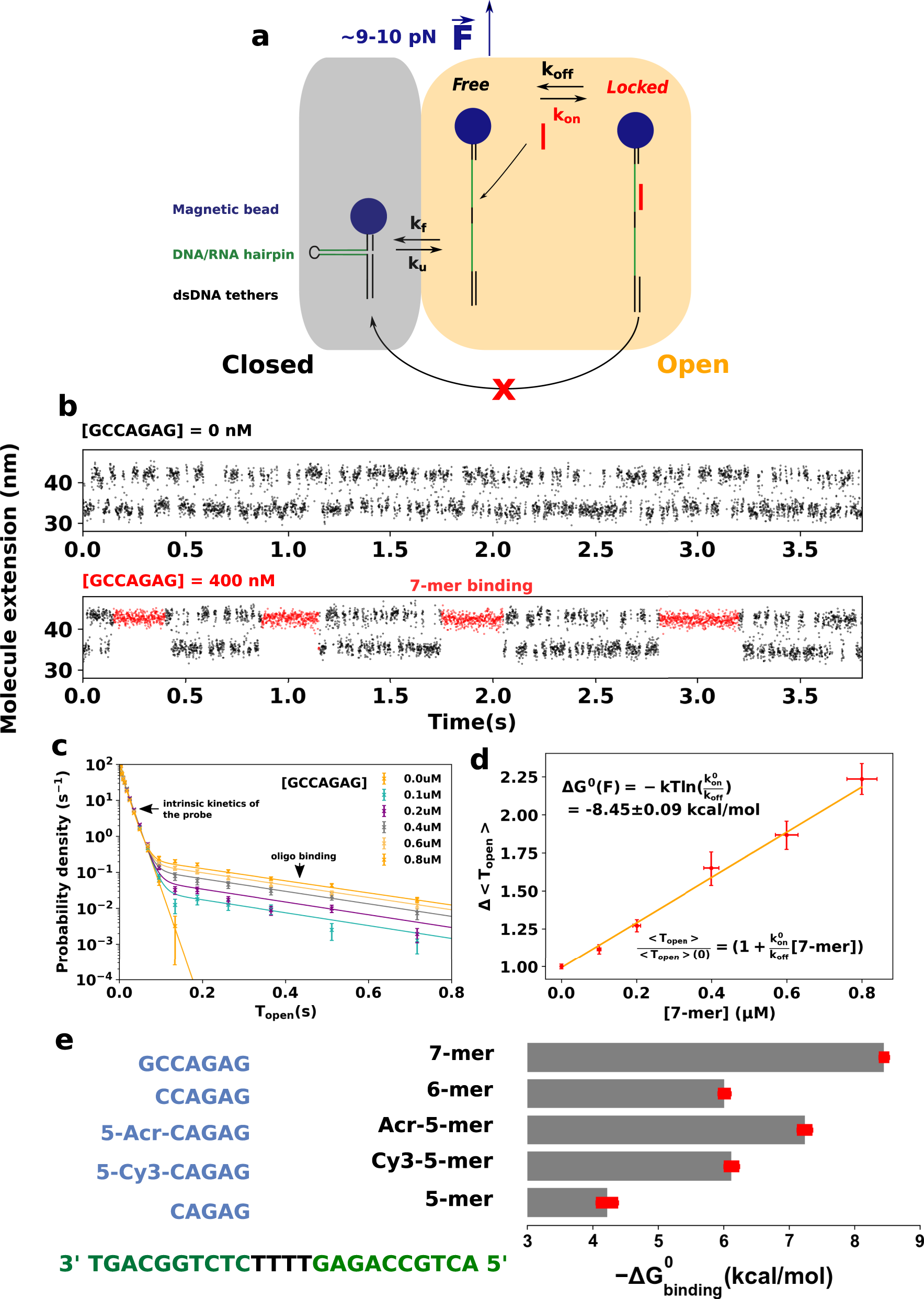
(a) When an oligonucleotide binds to the probe, it blocks it transiently in the open state. b Larger time view of the measured extension of the hairpin in the absence of oligonucleotide in solution (top) and in the presence of the complementary 7-mer (bottom). c Distribution of the times spent by the DNA probe in its open state as a function of the concentration of 7-mer. The clear double-exponential distribution allows us to deduce the binding rates kon and koff. d Relative evolution of the mean time spent by the DNA probe in the open state \(\overline{{{{{{T}}}}}_{{{{{{{{\rm{open}}}}}}}}}}\) as a function of the concentration of 7-mer. The slope allows us to compute the energy of binding. Y errors are computed through bootstrap resampling. X errors are based on a putative error of 5% on the concentration. Each data point consists of the averaging of at least 7000 events. e Free energies of binding of five different oligonucleotides measured with kinetic locking. The sequence of the probe is shown in green and the sequences of the oligonucleotides are in blue. Cy3 and acridine modifications increase substantially the binding energy. Errors are based on the covariance of the parameters inferred from the linear fits like the one shown in (d).
Detection of the nanometric extension change induced bu the hybridization of short oligonucleotides
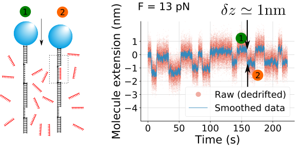
I measured the kinetic parameters of hybridization of short DNA oligonucleotides (7-10 nt) without the use of fluorescence. The goal is to tabulate thermodynamical data of such short segments without having to introduce fluorophores. When pulled at 13 pN, dsDNA is shorter than ssDNA. The difference of length for 8 nt is 1 nm. By detecting such extension change, I deduce the presence of the oligonucleotide and its kinetic parameters.
Measurement of binding/unbinding parameters of helicases
Helicases are molecular motors that walks ( translocates ), along nucleic acids, in order to unwind the duplex structure. I use the millisecond resolution of our magnetic tweezers (for 10 nm extension changes) in order to resolve the fluctuations of a 10 bp hairpin. When an helicase binds to the hairpin, it changes importantly the energetics of hybridization and thus the time spent by the latter in the closed state. We can thus infer from the kinetics of fluctuation the presence of the helicase. This technique does not need fluorescent labeling of proteins.
Characterization of helicase stepping
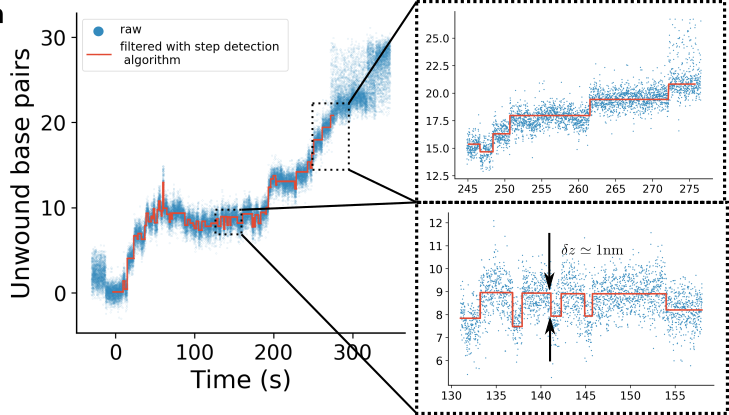
When an helicase translocates along a duplexed nucleic acid that is stretched at a force of 8-9 pN, each unwound base results in a change of extension of the nucleic acid of roughly 1 nm. At low ATP, some helicases with a strong grip on nucleic acids, like Upf1, are slowed down up to time scales that can be resolved by our system (0.5 second for 1 nm steps).
Magnetic tweezers
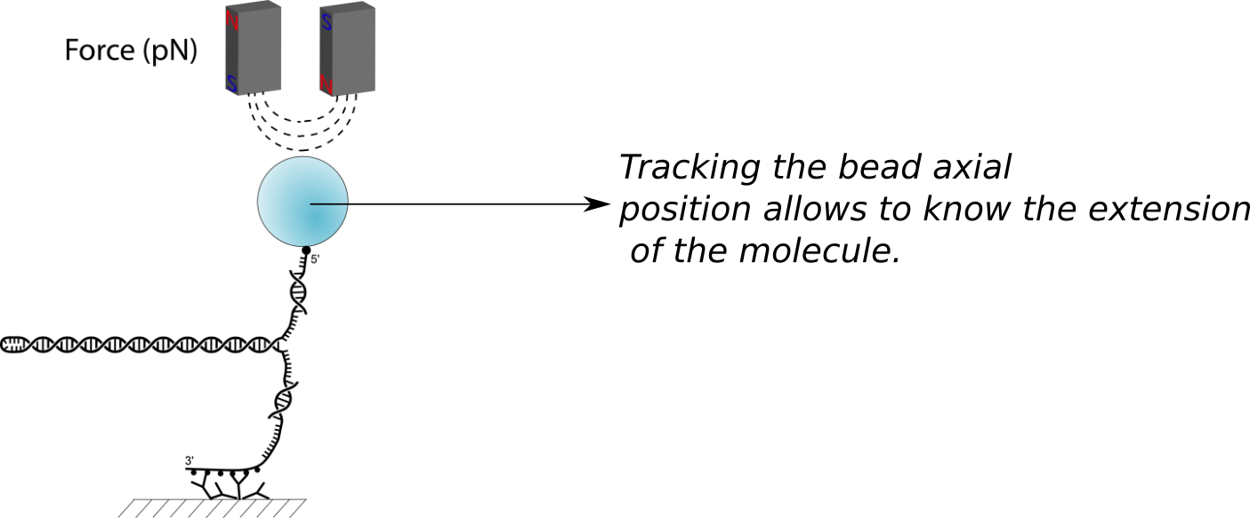
Magnetic tweezers are small magnets coupled to a precise 3D-optical detection systems that allow characterizing biopolymers. In short, the technique consists in tethering biomolecules such as proteins or nucleic acids to a glass surface on one side and to magnetic microspheres on the other. The magnets apply a force to the magnetic microspheres that is transmitted to the biomolecule of interest. The physical response of the biomolecule - its extension - is measured through the 3D-tracking of the position of the microsphere.
The technique is a powerful biophysical technique allowing :
- the assessment of the canonical thermodynamical properties of biomolecules with a much better precision than traditional calorimetric experiments.
- the validation of the underlying models of statistical physics.
- the direct and label-free observation of the interaction of molecular motors with nucleic acids or cell filaments. Molecular motors are proteins that perform specific tasks related to the structure of a long biopolymer, such as unwinding it (helicases), replicating it (polymerases), rearranging its folds and nodes (topoisomerases). The interaction of such molecular motors with a biopolymer strectched at a force of a few piconewtons gives often rise to a signature in extension that can be measured and analyzed.
Compared to other single-molecules micromanipulation techniques, such as optical trapping or atomic force miscrocopy (AFM), magnetic tweezers present several advantages :
- The force applied on the molecule is extremely stable, because its typical distance of variation (~200 micrometers) is much larger than the studied biopolymers (a few microns). Thus, a change of extension of the oberved molecule does not imply a significant change of force.
- The local temperature is also extremely stable, contrarely to the case of optical tweezers where thermal fluctuations arise from the energetic dissipation by the trapping laser.
- Rotating the magnets allows to study the effect of torsion on the molecules to be studied.
- Attachment is robust, allowing to measure properties of a unique molecule during several tens of hours.
- Most importantly, the technique is highly parallelizable , allowing to study hundreds of beads in parallel, while optical tweezers need one laser trap and AFM one cantilever per molecule.
However, measurements made with magnetic tweezers are known to be less precise . Optical tweezers are now able to resolve nanometric steps at a time scale of a few milliseconds , allowing the resolution of the discrete kinetics of molecular motors such as helicases and kinesins. However, the most precise signals measured with magnetic tweezers were until now extension changes of ~10 nm and were measured on time scales of ~10 s.
Increasing the precision
Spreading the PSF to reduce photon noise in spite of the camera welldepth
One of the source of noise in magnetic tweezers experiments is the photon noise : the less you collect photons at each frame, the more uncertainty on the position of the bead. The position of the bead is indeed encoded in the Point-Spread-Function (PSF) of the setup. A good sampling of this PSF is therefore necessary.
The most simple way to increase the number of photons received by unit of time to localize one bead is to increase the intensity of the incident light. However, the number of photons that a pixel of a camera can analyze in one frame is physically limited (welldepth of the camera, typically 30,000 photons). Increasing the intensity of the incident light thus implies increasing the acquisition frequency (nb of frames by second) of the camera in order to keep the number of photons received in each frame constant. Here again, this acquisition frequency is limited by the maximal transfer rate between the camera and the computer.
In order to increase the precision, a solution is to spread the PSF on more pixels in order to circumvent the obstacle of the welldepth. This is the goal of Stereo Darkfield Interferometry , a technique conceived by my advisor Vincent Croquette and developed by our team with Thibault Vieille and Gaël Radou .
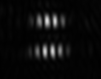
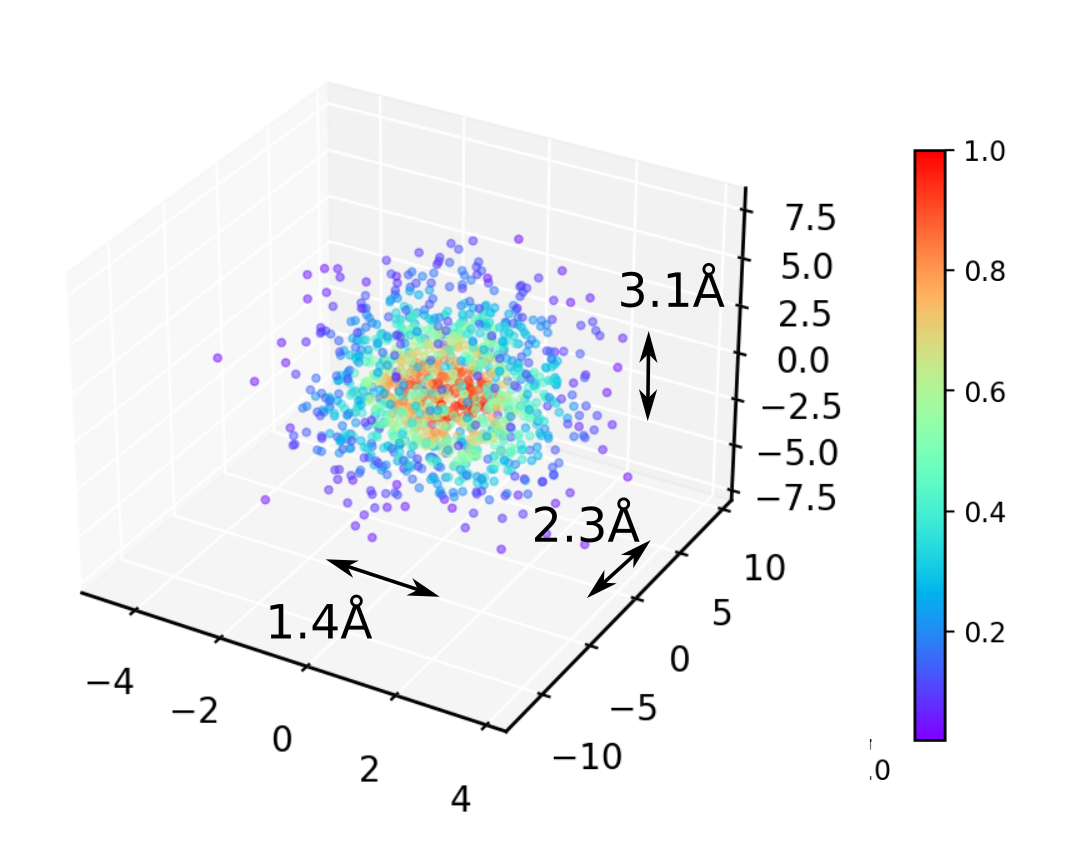
This new technique allows to reach 2 Angströms of optical noise (spread of localization distribution for a fixed object at maximum light intensity) in all three directions.
Correcting tilted optical axis
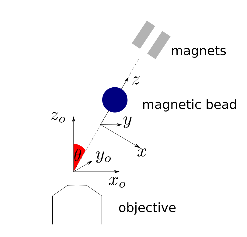
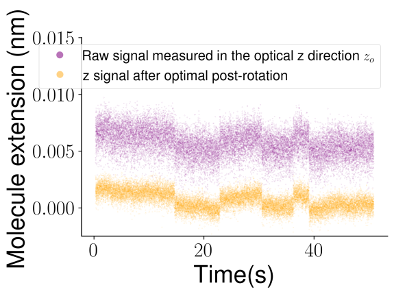
An other source of noise that is critical and can be easily corrected is a misalignment of the magnetic axis and the optical axis. Indeed, the Brownian motion of an overdamped microparticle is much larger in the direction perpendicular to the force. It is thus absolutely necessary to measure its displacement in the right direction. In order to get rid of possible misalignments (2° of tilt doubles the noise), I proposed to postprocess the signal by finding, by simple minimization, the axis along which the noise is at its smallest.
Towards better tethering and surface passivation
In order to reduce the Brownian motion of the bead, it is fundamental to work with stiffer and thus shorter molecules. Shorter molecules however induce a higher proximity to the surface. When a microspheres is very close from the surface as in our experiments (40 nm from the surfaces for a bead of radius 500 nanometers), two major problems arise :
- The proximity of the surface increases the probability of non specific interaction of the bead with the surface
- The proximity of the surface increases dramatically the effective viscosity of water, shifting the Brownian motion towards slower frequency and thus increasing the measured noise (by roughly a factor 4). This noise can not be reduced by using longer dsDNA handles as the decrease of viscosity is more than compensated by the decrease of stiffness of the polymers.